Request Quote
Scan the QR code below to connect with us on WhatsApp.

Request Quote
Scan the QR code below to connect with us on WhatsApp.

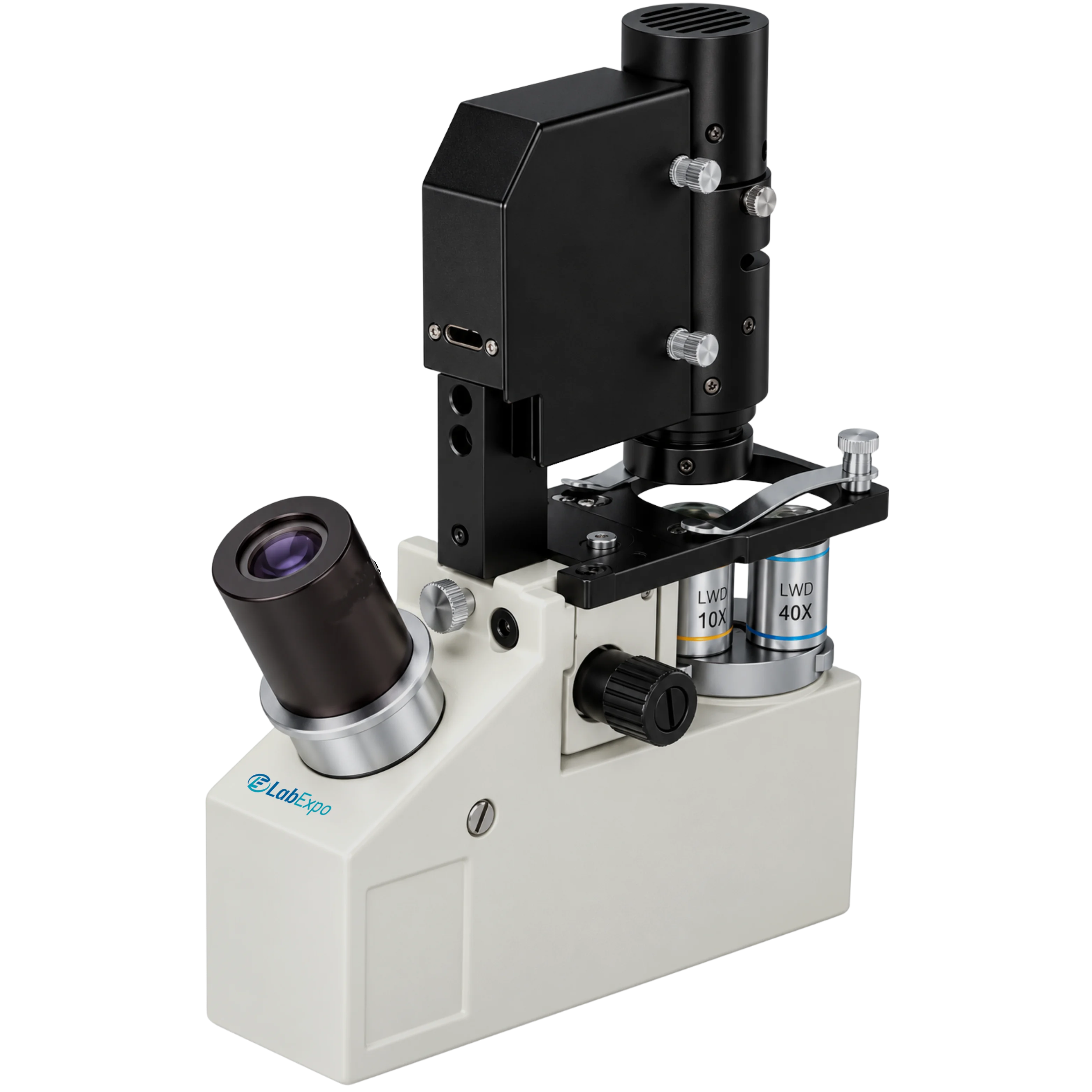
Lab Expo Inverted Biological Microscope 43-IBM400 is with compact & sleek design is equipped with stable & reliable ‘T’ type base, phase contrast long working distance objectives. It helps user to configure the cell more clearly thereby provides high quality images of the specimen. This user-friendly inverted microscope is used in many branches of biology.
$3,113.00 $ 2594
| Viewing head | Monocular viewing head |
| Eyepiece | 10X/ 18 mm |
| Objectives | 4X/N.A. 0.08, working distance= 16 10X/N.A. 0.25, working distance= 3.6 40X/N.A. 0.6, working distance= 0.6 |
| Nosepiece | Triple nosepiece 4X/ N.A. 0.8, WD 16 mm |
| Optical system | Finite optical system |
| Condenser | Bright field condenser with iris diaphragm |
| Stage dimensions | Plain stage with slide specimen clips |
| Range of vertical stroke | Up 1.6 , down 1 |
| Illumination | 0.1 W LED illumination (rechargeable) |
| Dimension | 200 × 200 × 80 mm |
| Gross weight | 2 kg |
Function Implementation Cell culture labs 10X/ N.A. 0.25, working distance= 3.6 enables observation of live specimens in flasks Microbiology workflows Triple nosepiece 4X/ N.A. 0.8, WD 16 mm supports rapid objective switching Quality control stations Plain stage with slide specimen clips secures samples during routine inspection Research benchtop use Dimension: 200 × 200 × 80 mm fits space-constrained laboratory setups Field sampling units Gross weight: 2 kg allows transport between collection sites and lab analysis
Lab Expo Inverted Biological Microscopes are structured around inverted optical geometry to enable direct observation of cells and cultures within vessels. A rigid T-base frame minimizes vibration, while long working-distance phase contrast objectives and matched condenser systems support clear imaging of thick samples. Binocular or trinocular viewing options, precision focus control, and stable illumination ensure accurate, repeatable cell analysis for research, clinical, and academic laboratory workflows.


