Request Quote
Scan the QR code below to connect with us on WhatsApp.

Request Quote
Scan the QR code below to connect with us on WhatsApp.
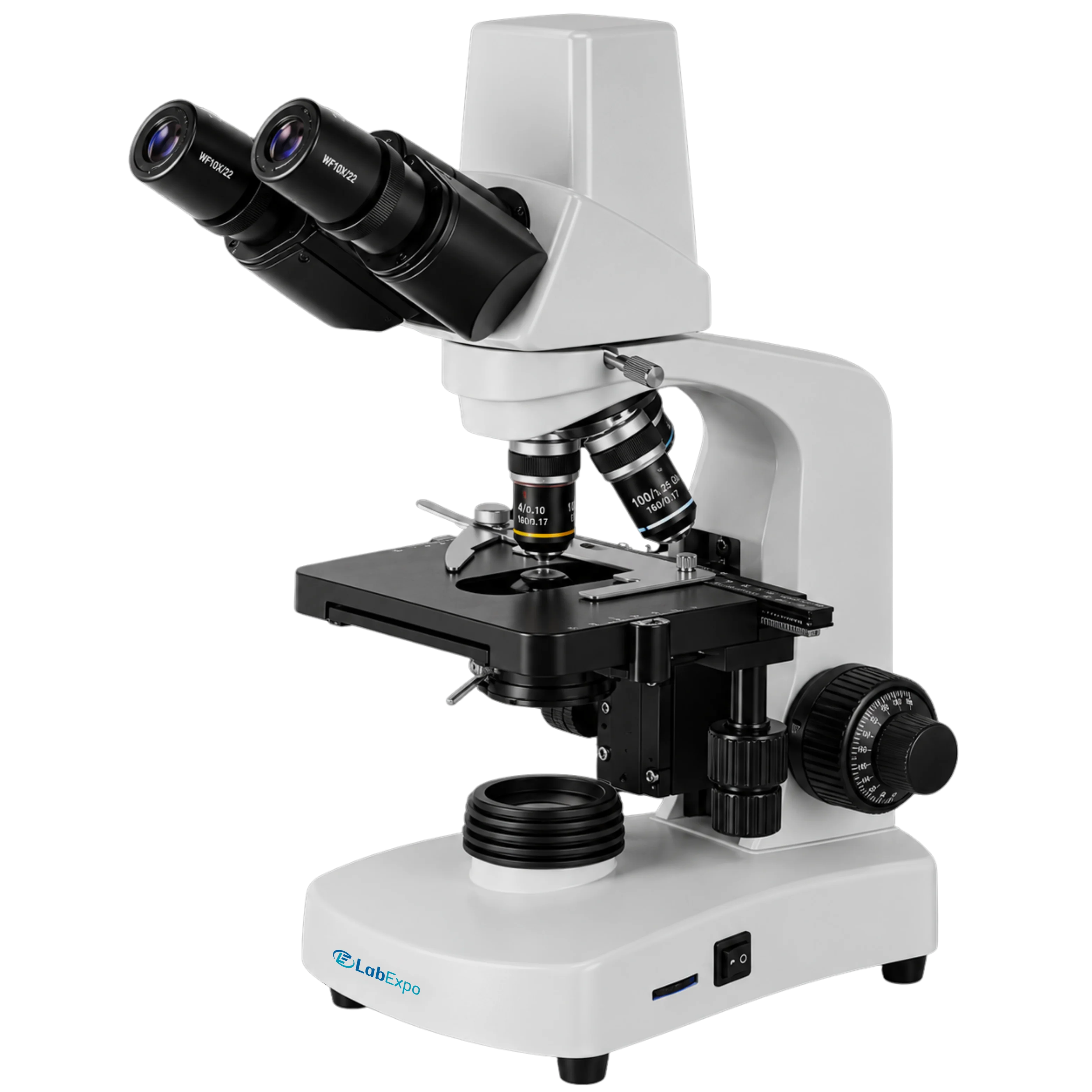

Lab Expo Digital Microscope 43-DMS300 features built-in USB camera, and PC/monitor attachment with professional software offers high resolution images. Digital microscopes allows one to view the sample without straining the eyes. Coaxial coarse and fine focusing adjustment of this microscope gives sharp and clear image.
$2,364.00 $ 1970
| Viewing head | Compensation free binocular head inclined at 30° , interpupillary distance 48 -75 mm Range of viewing field: 90% |
| Eyepiece | WF10X/18 mm |
| Objectives | Achromatic objective: 4x, 10x, 40x(s), 100x(s, oil) |
| Stage | Double layer mechanical stage: 132×142 mm Moving range: 75×40 mm |
| Condenser | Abbe N.A. 1.20 with iris diaphragm and filter |
| Focusing | Coaxial coarse and fine adjustment system Range 24 mm, fine division 0.004 mm |
| Nosepiece | Backward quadruple nosepiece |
| Illumination | S-LED brightness adjustable Halogen lamp 6V/20W with adjustable brightness |
| Output | USB 2.0 |
| Operating system | Windows 2000/XP/Vista |
| Software | Camea software: Scope image 9.0 |
| Valid Pixel | Camera Valid pixel: 1280×1024 (1.3 mega pixel) |
| Dimension | 395×275×510 mm |
| Net weight | 8 kg |
| Gross weight | 9 kg |
Function Implementation Quality control labs 1280×1024 pixel resolution enables detailed sample inspection Clinical diagnostics 100x oil immersion objective supports cellular structure analysis Educational institutions WF10X/18 mm eyepiece allows comfortable extended viewing Research facilities 75×40 mm stage movement range facilitates precise specimen positioning Industrial material testing Coaxial fine adjustment 0.004 mm ensures accurate focus calibration
| Accessories No. | Name |
| 1 | Eyepiece: P16X/11 mm |
| 2 | Phase contrast kit |
| 3 | Dark field attachment |
| 4 | Polarization attachment |
Lab Expo Digital Microscopes are structured around integrated optical–digital imaging paths to deliver high-resolution visualization on a built-in LCD display. Precision coarse and fine focus control, adjustable LED illumination, and plan achromatic optics ensure sharp image formation across specimen types. USB connectivity, Android compatibility, and intelligent analysis software support measurement, documentation, and efficient data handling for research, industrial inspection, education, and clinical laboratory workflows.


